L’ wave in echocardiogram
‘L’ Wave in Echocardiography – Explained
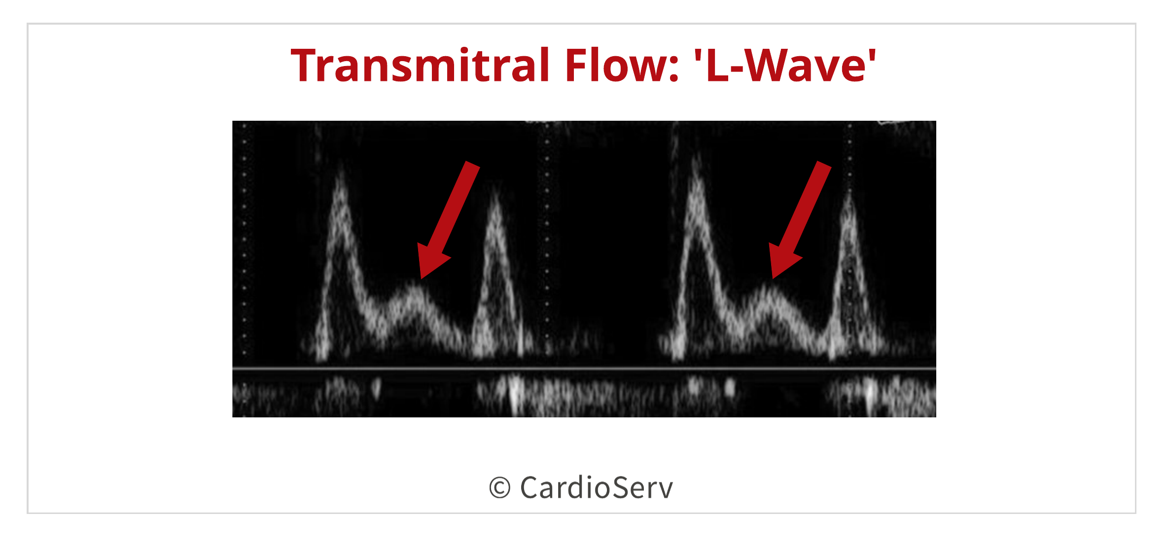
The ‘L’ wave is a mid-diastolic transmitral flow signal seen on pulsed-wave Doppler imaging of the mitral inflow in certain pathological conditions.
What is the ‘L’ wave?
- It is an additional diastolic wave between the E wave (early filling) and A wave (atrial contraction) on mitral inflow Doppler.
- Appears after the E wave but before the A wave during mid-diastole.
- It reflects mid-diastolic mitral inflow due to persistent or increased left atrial pressure gradient across the mitral valve during diastole.
Echocardiographic Appearance
- Detected in apical 4-chamber view with pulsed-wave Doppler at the mitral leaflet tips.
- The Doppler flow pattern shows:
E wave → L wave → A wave
Conditions Associated with L Wave
| Condition | Explanation |
|---|---|
| Diastolic Dysfunction (Grade 2 or 3) | Impaired relaxation with elevated LV filling pressures |
| Advanced Heart Failure | With elevated left atrial pressure and pseudonormal/restrictive filling |
| Atrial Fibrillation | Seen due to loss of A wave and compensatory filling |
| Post-cardiac transplant | Can occur due to abnormal diastolic compliance |
| Constrictive Pericarditis or Restrictive Cardiomyopathy | Due to rapid early filling and stiff ventricles |
Mechanism Behind the L Wave
- In mid-diastole, there’s usually a pause in mitral flow.
- However, in certain diseases, LA pressure remains elevated, causing persistent flow across the mitral valve even in mid-diastole.
- This causes an extra wave—the L wave—to appear.
Clinical Significance
- Marker of elevated left ventricular filling pressure.
- May suggest adverse prognosis in heart failure.
- Useful in non-invasive grading of diastolic dysfunction.
Mnemonic: “EL-A” pattern
Early diastolic
Late-mid diastolic
Atrial contraction
(E → L → A)
1. What is the L wave in mitral inflow Doppler?
- It is a mid-diastolic flow signal seen between the E and A waves.
- Detected on pulsed-wave Doppler at the mitral leaflet tips.
- It appears due to persistent pressure gradient from LA to LV in mid-diastole.
- It is often observed in pathological diastolic filling conditions.
- The pattern seen is E-L-A (Early, Mid, and Atrial filling waves).
2. What are the conditions associated with the presence of an L wave?
- Grade 2 and Grade 3 diastolic dysfunction.
- Advanced heart failure with raised LV filling pressure.
- Atrial fibrillation (absence of A wave makes L wave more visible).
- Restrictive cardiomyopathy and constrictive pericarditis.
- Post-cardiac transplant due to altered compliance.
3. What does the presence of an L wave indicate clinically?
- It suggests elevated left ventricular filling pressures.
- Associated with abnormal diastolic function.
- May reflect reduced LV compliance.
- Indicates worse prognosis in some heart failure patients.
- Helps grade diastolic dysfunction in echocardiography.
4. How is the L wave detected on echocardiography?
- Use pulsed-wave Doppler at the mitral leaflet tips.
- Typically assessed in the apical 4-chamber view.
- Appears between the early (E) and late (A) diastolic waves.
- Seen as a distinct mid-diastolic positive wave.
- Requires slow heart rate or high filling pressure to be clearly visualized.
5. What are the hemodynamic mechanisms that lead to L wave formation?
- Persistent pressure gradient between LA and LV during mid-diastole.
- High LA pressure sustains flow across mitral valve.
- Rapid early filling followed by a brief mid-diastolic flow pause.
- Then mid-diastolic LA pressure rises again, causing another wave (L wave).
- Reflects altered ventricular relaxation or stiffness.
6. How does atrial fibrillation affect L wave visibility?
- A wave is absent due to lack of atrial contraction.
- Makes L wave more prominent and easier to detect.
- E-L pattern replaces the usual E-A pattern.
- Seen in AF patients with elevated LA pressure.
- Useful in evaluating diastolic function in AF.
7. What is the timing of the L wave during the cardiac cycle?
- Occurs during mid-diastole.
- Appears after the E wave but before the A wave.
- Usually around 150–200 milliseconds after E wave.
- Detected between rapid early filling and atrial contraction.
- Only visible when diastolic filling duration is sufficient.
8. What is the prognostic significance of the L wave?
- Associated with elevated left-sided filling pressures.
- Suggests more advanced diastolic dysfunction.
- May predict worse outcomes in heart failure patients.
- Indicates impaired ventricular relaxation and compliance.
- Disappearance may reflect successful therapy.
9. How does heart rate affect the appearance of the L wave?
- Bradycardia enhances its visibility.
- Prolonged diastole separates E, L, and A waves clearly.
- Tachycardia may fuse E and L waves, making L wave undetectable.
- Very fast rates can eliminate mid-diastolic flow.
- Best detected in slow to moderate heart rates.
10. What is the significance of the E-L-A pattern on mitral Doppler?
- Helps in grading and identifying advanced diastolic dysfunction.
- It indicates presence of L wave between E and A waves.
- Suggests abnormal diastolic filling dynamics.
- Seen in restrictive or pseudonormal filling patterns.
- Reflects elevated LA pressure during all phases of diastole.
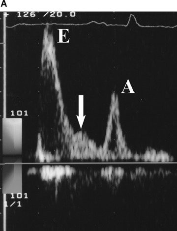
Image Question-56 – Medicine Question Bank
L wave in echocardiography refers to a mid-diastolic flow seen in pulse wave Doppler and M-mode echocardiography.
L-wave.png
Clinical Significance:The presence of an L wave can be an indicator of diastolic dysfunction and elevated left ventricular filling pressures, while its disappearance can suggest successful treatment and improved heart function.
Types of L- Wave in Echocardiogram
Two types of L-waves.
Type I
Distinct L-wave (Type I) with an accelerating and decelerating portion.
Type II
Merged L-wave (Type II) having only a decelerating portion, manifesting as an abrupt change in the deceleration segment of the flow profile


