Frog sign
Frog sign
Frog sign is seen in case of –
A. Atrial Fibrillation
B. Atrial flutter
C. AVNRT
D. AVRT
What is the mechanism of Frog Sign?
AVNRT – There is AV dissociation
Canon A waves -occur with AV dissociation result from simultaneous contractions of the atria and ventricles against closed mitral and tricuspid valves
Canon A waves – cause reflux of blood into the neck veins.
1. What does the “Frog Sign” indicate in a patient with PSVT?
A. Bradycardia due to sinus node dysfunction
B. Atrial flutter with variable block
C. Cannon A waves due to AV dissociation
D. Pulmonary hypertension signs
Explanation: The Frog Sign is due to cannon A waves caused by atrial contraction against a closed tricuspid valve during AVNRT.
2. Frog Sign is most commonly seen in which type of PSVT?
A. Atrial fibrillation
B. AVNRT (Atrioventricular Nodal Reentrant Tachycardia)
C. WPW syndrome with orthodromic conduction
D. Sinus tachycardia
Explanation: AVNRT is associated with AV dissociation during tachycardia, causing visible cannon A waves—termed Frog Sign.
3. Cannon A waves are produced when:
A. Atria contract against a closed tricuspid valve
B. Ventricle contracts against closed aortic valve
C. Tricuspid regurgitation is present
D. Ventricular diastole is prolonged
Explanation: The atrium contracting when the tricuspid valve is closed leads to retrograde venous pressure—seen as cannon A waves.
4. Frog sign is best observed in:
A. Carotid artery
B. Jugular venous pulse
C. Radial artery
D. Apex impulse
Explanation: Jugular veins reflect right atrial activity. Frog sign manifests as giant venous pulsations here.
5. Which physical description best matches the frog sign?
A. Bulging neck veins resembling frog’s throat
B. Weak thready pulse in extremities
C. Pulsatile liver edge
D. Cyanotic lips with peripheral edema
Explanation: The term “Frog Sign” comes from the bulging of neck veins during episodes of PSVT resembling a frog’s throat.
6. Which of the following triggers is commonly associated with AVNRT and Frog Sign?
A. Hypercalcemia
B. Hypothyroidism
C. Caffeine or emotional stress
D. Chronic beta-blocker use
Explanation: AVNRT and Frog Sign are often seen in young people triggered by stress, caffeine, or excitement.
7. The mechanism of AVNRT involves:
A. Accessory pathway between atria and ventricles
B. Reentry circuit within AV node
C. Automatic focus in the pulmonary veins
D. SA node conduction delay
Explanation: AVNRT is caused by a reentrant loop within the AV node using slow and fast pathways.
8. Which ECG feature supports AVNRT diagnosis?
A. Delta wave
B. Retrograde P wave just after QRS
C. Multiple P waves for each QRS
D. Irregularly irregular rhythm
Explanation: In AVNRT, retrograde P waves may be buried or just after QRS due to simultaneous atrial and ventricular contraction.
9. What is the heart rate usually observed in AVNRT?
A. 40–60 bpm
B. 150–250 bpm
C. 90–100 bpm
D. 60–70 bpm
Explanation: AVNRT typically presents with a narrow-complex tachycardia at a rate between 150–250 bpm.
10. Frog sign is absent during:
A. Normal sinus rhythm
B. AVNRT episode
C. Atrial tachycardia
D. Ventricular ectopy
Explanation: Cannon A waves—and thus the frog sign—occur only during arrhythmias involving AV dissociation, not in normal rhythm.
11. The “cannon A wave” is seen due to:
A. Atrial contraction during ventricular systole
B. Atrial fibrillation
C. SA node pause
D. Mitral stenosis
Explanation: Cannon A waves occur when atria contract while the tricuspid valve is still closed, usually during AV dissociation.
12. AV dissociation means:
A. Atria and ventricles contracting independently
B. SA node suppression
C. AV node block only
D. Simultaneous atrial and ventricular contraction always
Explanation: AV dissociation occurs when atrial and ventricular contractions are no longer synchronized.
13. In what clinical setting is the Frog Sign most easily observed?
A. Patient at rest, supine
B. During PSVT episode while sitting upright
C. During sleep
D. After exercise recovery
Explanation: Frog sign becomes visible during PSVT episodes due to jugular venous pulsation when sitting or semi-reclined.
14. Which vein best shows the Frog Sign?
A. Internal jugular vein
B. Subclavian vein
C. Basilic vein
D. Cephalic vein
Explanation: The internal jugular vein lies in line with right atrial pressure and best reflects atrial activity.
15. Termination of AVNRT and Frog Sign is often achieved by:
A. Epinephrine infusion
B. Vagal maneuvers or adenosine
C. Beta agonists
D. Digoxin bolus
Explanation: Vagal maneuvers or adenosine transiently block the AV node and terminate AVNRT, resolving the Frog Sign.
16. What physical exam maneuver can help accentuate the Frog Sign?
A. Carotid massage
B. Inspiration
C. Sitting upright with neck extended
D. Coughing vigorously
Explanation: The Frog Sign becomes more visible in the upright position with neck extended, allowing jugular pulsations to be seen.
17. Which of the following is NOT a feature of AVNRT?
A. Sudden onset and offset
B. Narrow QRS complexes
C. Irregularly irregular rhythm
D. Retrograde P waves
Explanation: AVNRT typically produces a regular rhythm. Irregularly irregular rhythms are characteristic of atrial fibrillation.
18. Frog Sign may resemble pulsations seen in:
A. Mitral valve prolapse
B. Complete heart block
C. Aortic dissection
D. Restrictive cardiomyopathy
Explanation: Cannon A waves are also seen in complete heart block, due to AV dissociation similar to AVNRT.
19. What diagnostic test confirms the presence of AVNRT in a patient with Frog Sign?
A. 12-lead ECG during tachycardia
B. Chest X-ray
C. Transthoracic echocardiogram
D. Holter during normal sinus rhythm
Explanation: A 12-lead ECG during the PSVT episode confirms the diagnosis of AVNRT by showing narrow QRS tachycardia with hidden or retrograde P waves.
20. A patient presents with visible neck pulsations and palpitations. What is the next best step?
A. Obtain ECG to assess for PSVT
B. Schedule routine echocardiogram
C. Start long-term anticoagulation
D. Reassure the patient without workup
Explanation: ECG during symptoms is essential to confirm PSVT, especially when Frog Sign is observed.
Frog sign
| No. | Frog sign – Key Point |
|---|---|
| 1 | Frog Sign refers to visible pulsations in the neck veins during PSVT. |
| 2 | It is caused by “cannon A waves” due to atrial contraction against closed tricuspid valve. |
| 3 | Most commonly seen in AVNRT, a type of paroxysmal supraventricular tachycardia. |
| 4 | The sign is transient and occurs only during tachycardia episodes. |
| 5 | Jugular venous pulse is the best location to observe the Frog Sign. |
| 6 | AV dissociation is the underlying mechanism of cannon A waves. |
| 7 | Simultaneous atrial and ventricular contraction causes the backflow into neck veins. |
| 8 | Neck pulsations resemble the bulging throat of a frog—hence the name. |
| 9 | ECG during symptoms shows narrow complex tachycardia with hidden/retrograde P waves. |
| 10 | Typical AVNRT rate is 150–250 bpm. |
| 11 | Frog Sign disappears when normal sinus rhythm is restored. |
| 12 | Can be triggered by stress, caffeine, or vagal tone changes. |
| 13 | Seen more clearly in upright or semi-recumbent position. |
| 14 | Best visualized in the internal jugular vein, not the carotid artery. |
| 15 | May resemble cannon waves of complete heart block but occurs with tachycardia. |
| 16 | Diagnosis is confirmed with ECG during symptoms. |
| 17 | Management includes vagal maneuvers or adenosine to terminate the arrhythmia. |
| 18 | Carotid sinus massage may abort AVNRT and stop the Frog Sign. |
| 19 | Frog Sign is a classic teaching example of a visible clinical sign of arrhythmia. |
| 20 | It is an important bedside clue for recognizing AVNRT in acute settings. |
1. What is the Frog Sign and in what context is it observed?
- Frog Sign refers to visible neck pulsations resembling a frog’s throat.
- It results from cannon A waves in the jugular venous pulse.
- Occurs when the atria contract against a closed tricuspid valve.
- Most commonly seen in AVNRT, a type of PSVT.
- Appears only during tachycardia episodes, not in normal rhythm.
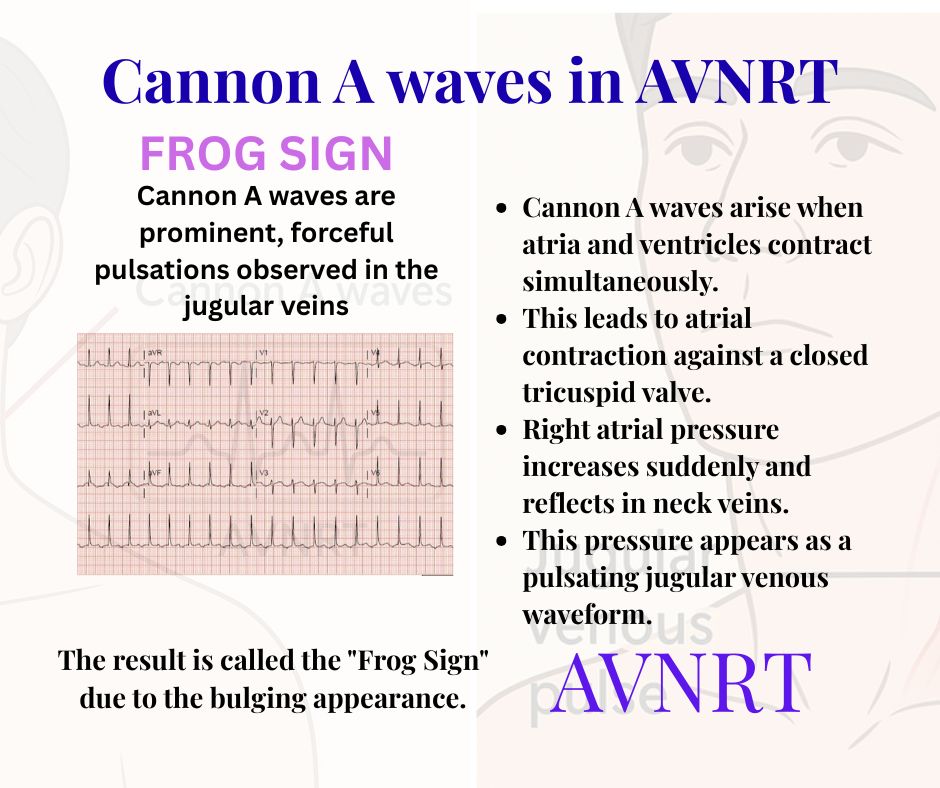
2. Explain the mechanism behind cannon A waves in AVNRT.
- Cannon A waves arise when atria and ventricles contract simultaneously.
- This leads to atrial contraction against a closed tricuspid valve.
- Right atrial pressure increases suddenly and reflects in neck veins.
- This pressure appears as a pulsating jugular venous waveform.
- The result is called the “Frog Sign” due to the bulging appearance.
3. Describe the ECG findings in AVNRT where Frog Sign is present.
- Narrow QRS complex tachycardia is seen.
- P waves may be hidden in or just after the QRS complex.
- Regular rhythm at a rate of 150–250 bpm.
- Retrograde P waves may appear in inferior leads (II, III, aVF).
- No visible PR interval or distinct P waves before QRS.
4. List conditions other than AVNRT where cannon A waves can occur.
- Complete heart block (3rd-degree AV block).
- Ventricular tachycardia with AV dissociation.
- Junctional rhythm without retrograde conduction.
- Premature ventricular contractions (PVCs).
- Pacemaker-induced AV dissociation.
5. What are some bedside techniques to visualize the Frog Sign?
- Position the patient semi-upright (30–45 degrees).
- Ensure good lighting on the neck area.
- Ask the patient to look to the left to expose the right IJV.
- Look for rhythmic, large pulsations synchronous with heartbeat.
- Observe during a tachycardia episode for maximal effect.
6. What is the clinical importance of recognizing Frog Sign?
- It serves as a physical clue to AVNRT or AV dissociation.
- Helps differentiate from other types of tachycardia.
- Suggests a reentrant mechanism involving AV node.
- Prompts immediate ECG confirmation and management.
- Provides a rapid bedside clue, especially in emergency settings.
7. How is AVNRT managed when Frog Sign is present?
- Initiate vagal maneuvers (Valsalva, carotid massage).
- If ineffective, give IV adenosine rapidly.
- Beta-blockers or calcium channel blockers may be used.
- In unstable patients, synchronized cardioversion is preferred.
- Long-term: consider catheter ablation of the slow AV nodal pathway.
8. How does Frog Sign help in differentiating AVNRT from atrial fibrillation?
- Frog Sign suggests regular AVNRT, not irregular AF.
- In AF, no organized atrial contraction occurs (no cannon waves).
- Jugular pulsations in AF are chaotic or absent.
- ECG in AF shows irregular rhythm with no distinct P waves.
- Visible neck pulsations during PSVT lean toward AVNRT.
9. What triggers can precipitate AVNRT and the appearance of Frog Sign?
- Emotional stress and anxiety.
- Excessive intake of caffeine or alcohol.
- Sudden postural changes or exertion.
- Increased vagal tone or vagal withdrawal.
- Underlying dual AV nodal pathways in the heart.
10. What are the limitations of using the Frog Sign for diagnosis?
- Frog Sign is transient and only visible during tachycardia.
- May be misinterpreted as carotid artery pulsation.
- Not all AVNRT cases show visible neck pulsations.
- Requires skilled observation and patient positioning.
- ECG is still essential for definitive diagnosis.
Cannon waves
Cannon waves occur when Atria contracts against a closing tricuspid valve of right ventricle .( There would be a equivalent left atrial cannon which goes into pulmonary vein as well


